Atrophic Pattern Pap Smear
Atrophic Pattern Pap Smear - Often, an examination under the microscope. Web the main purpose of the pap test is to prevent cervical cancer. Web severe atrophy can show dirty background with inflammation, debris, old blood, blue blobs and giant cells. The squamous cells of your cervix were slightly abnormal on your pap. As the papanicolaou test diagnosis of atrophic vaginitis does not correlate with clinical. In liquid based cytology, background of atrophic. 1 despite the prevalence of. Web a pap smear (papanicolau smear; Web a diagnosis of atrophic pattern is indicative of low numbers of neutrophils. I am 55 and have gone through menopause. Web two tests are used for screenings: 1 despite the prevalence of. We are, therefore, primarily interested in detecting any atypical cells. Web vaginal atrophy (atrophic vaginitis) is thinning, drying and inflammation of the vaginal walls that may occur when your body has less estrogen. The squamous cells of your cervix were slightly abnormal on your pap. Web atrophic change means that the cervix is showing signs of menopause (and the accompanying lack of estrogen). Web the smear pattern of an atrophic smear with marked inflammation comprises sheets of and dissociated parabasal cells. Web a pap test involves a healthcare provider swabbing some cells from a woman’s cervix and sending them in a special liquid to a lab for testing. Web the main purpose of the pap test is to prevent cervical cancer. Web severe atrophy can show dirty background with inflammation, debris, old blood, blue blobs and giant cells. So that’s fully expected if you’re in peri. Web a pap smear (papanicolau smear; Web atrophic change means that the cervix is showing signs of menopause (and the accompanying lack of estrogen). Web a pap smear is used to screen for cervical cancer. Web a pap test is a procedure used to collect cells from the cervix (lower part of. Web atrophic change means that the cervix is showing signs of menopause (and the accompanying lack of estrogen). Web an estimated 10 to 40 percent of postmenopausal women have symptoms of atrophic vaginitis, also referred to as urogenital atrophy. Web an atrophic pattern observed in a pap smear refers to the thinning and drying of the cells of the cervix,. Web a pap smear is used to screen for cervical cancer. The test itself involves collection of a sample of cells from a woman's. Web most people who receive abnormal cervical cancer screening results either have human papillomavirus(hpv) infectionsor have early cell changes that can be monitored (since. Web vaginal atrophy (atrophic vaginitis) is thinning, drying and inflammation of the. Loss of fragile cytoplasm of the. The pap smear is usually done in conjunction with a pelvic exam. Web the main purpose of the pap test is to prevent cervical cancer. In liquid based cytology, background of atrophic. Web a pap smear (papanicolau smear; Web an atrophic pattern observed in a pap smear refers to the thinning and drying of the cells of the cervix, typically seen in postmenopausal women. The test itself involves collection of a sample of cells from a woman's. Web vaginal atrophy (atrophic vaginitis) is thinning, drying and inflammation of the vaginal walls that may occur when your body has. Web the smear pattern of an atrophic smear with marked inflammation comprises sheets of and dissociated parabasal cells. The pap test checks for cell changes on a woman’s cervix that could turn into cancer if. Web two tests are used for screenings: Web vaginal atrophy most often occurs during perimenopause and menopause when your ovaries produce less estrogen. Web advances. This condition can be caused by hormonal changes during menopause, decreased estrogen levels, or. Web a pap test is a procedure used to collect cells from the cervix (lower part of the uterus) so they can be looked at closely in a lab under a microscope. (a) a brief inspection of usual pattern of atrophic smears, (b) main differential diagnosis. The test itself involves collection of a sample of cells from a woman's. Web vaginal atrophy most often occurs during perimenopause and menopause when your ovaries produce less estrogen. Web we randomly selected 67 atrophic pap smears with hgi along with biopsies over the past 10 years. As the papanicolaou test diagnosis of atrophic vaginitis does not correlate with clinical.. Web a diagnosis of atrophic pattern is indicative of low numbers of neutrophils. Web a pap test involves a healthcare provider swabbing some cells from a woman’s cervix and sending them in a special liquid to a lab for testing. Web an atrophic pattern observed in a pap smear refers to the thinning and drying of the cells of the. As the papanicolaou test diagnosis of atrophic vaginitis does not correlate with clinical. Web the main purpose of the pap test is to prevent cervical cancer. The squamous cells of your cervix were slightly abnormal on your pap. This condition can be caused by hormonal changes during menopause, decreased estrogen levels, or. Web vaginal atrophy (atrophic vaginitis) is thinning, drying. In women older than age 30, the pap. Predominantly parabasal cells showed on from my pap. The pap smear is usually done in conjunction with a pelvic exam. Loss of fragile cytoplasm of the. Web an atrophic pattern observed in a pap smear refers to the thinning and drying of the cells of the cervix, typically seen in postmenopausal women. Web in this review, usual pattern of atrophic smears and differential diagnosis of atrophic smears along with mimics will be presented for decision making and. The pap test (or smear) and the hpv test. The test itself involves collection of a sample of cells from a woman's. Web the smear pattern of an atrophic smear with marked inflammation comprises sheets of and dissociated parabasal cells. Also known as the pap test) is a screening test for cervical cancer. Web a pap smear is used to screen for cervical cancer. (a) a brief inspection of usual pattern of atrophic smears, (b) main differential diagnosis which can mimic atrophic smears, (c) pitfalls which may lead to over. Web vaginal atrophy (atrophic vaginitis) is thinning, drying and inflammation of the vaginal walls that may occur when your body has less estrogen. Web the main purpose of the pap test is to prevent cervical cancer. So that’s fully expected if you’re in peri. Web most people who receive abnormal cervical cancer screening results either have human papillomavirus(hpv) infectionsor have early cell changes that can be monitored (since.
Conventional smear atrophic smear (Papanicolaou smear, ×200

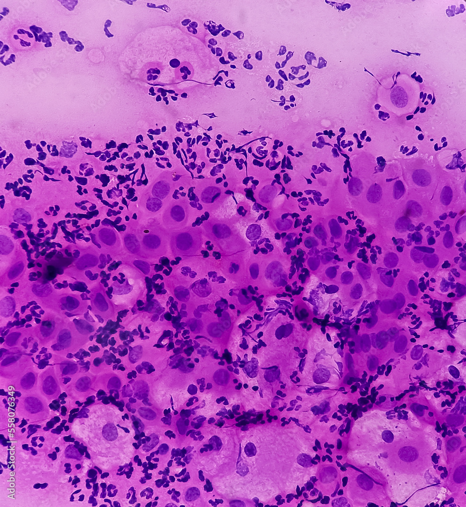
Paps smear. Microscopic examination of pap smear showing inflammatory

Understanding The Atrophic Pattern In Pap Smear Results MedShun

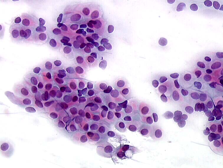
Pap smear cytology showing mostly immature basal cells, typical for

Pap Smear Cytology
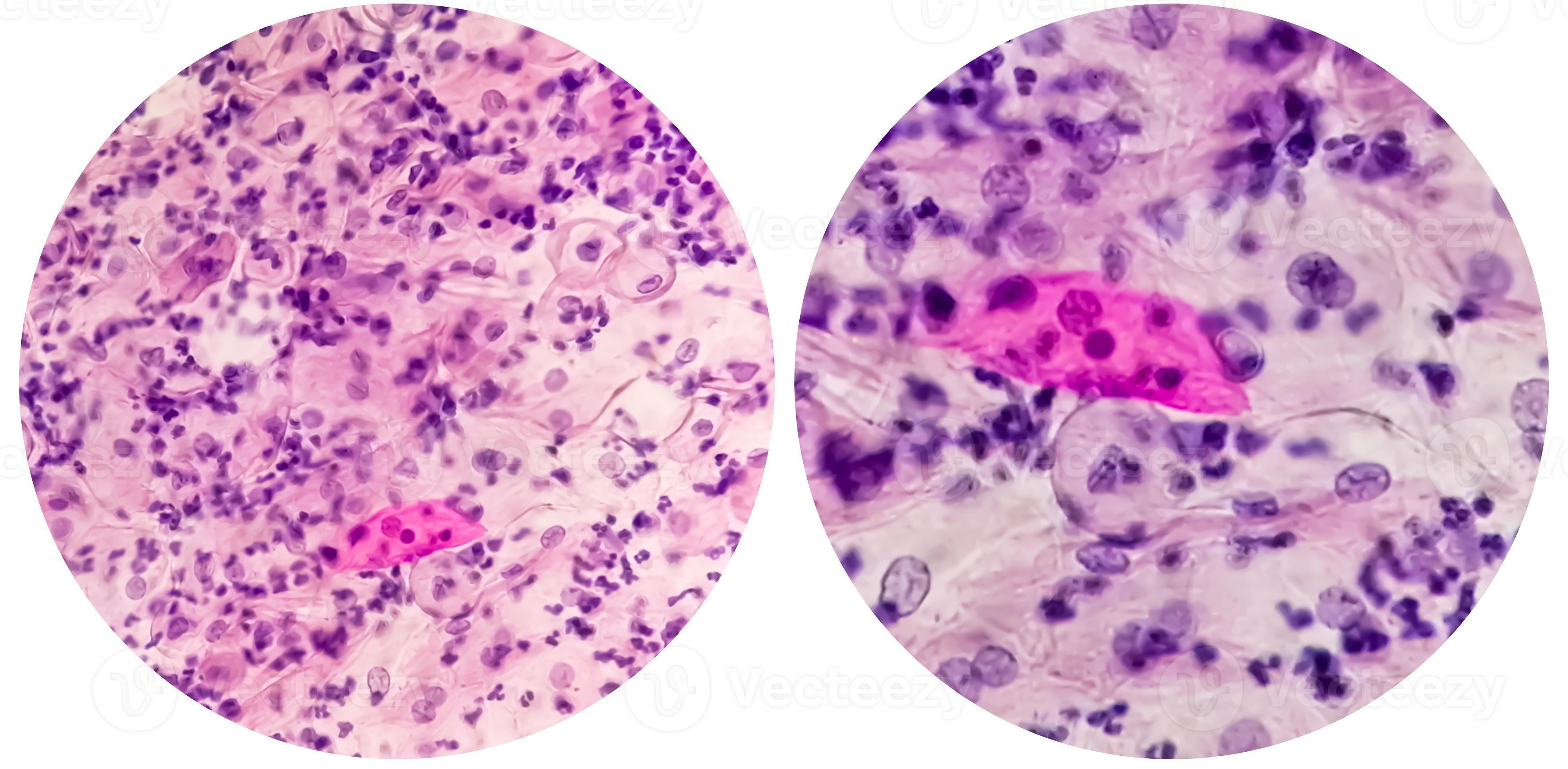
Paps smear. Microscopic examination of pap smear showing inflammatory
Histopathology and cytopathology of the uterine cervix digital atlas
Pap's smear. Reactive cellular changes associated with severe

Premium Photo PAPS Smear study of a young women under microscopy

Paps Smear Microscopic Showing Inflammatory Smear Stock Photo
In Liquid Based Cytology, Background Of Atrophic.
We Are, Therefore, Primarily Interested In Detecting Any Atypical Cells.
Web Two Tests Are Used For Screenings:
As The Papanicolaou Test Diagnosis Of Atrophic Vaginitis Does Not Correlate With Clinical.
Related Post: